Bioengineers use MRI to better understand cerebral palsy over time
03 February 2021
Auckland Bioengineering Institute (ABI) researchers are collaborating with Mātai, a medical imaging research centre in Tairāwhiti Gisborne, on what is possibly a world-first study that will help us understand more about cerebral palsy and how it affects muscle growth.

Using state-of-the-art MRI technology, the researchers will be looking at the inside of the legs of participants with cerebral palsy (CP) and comparing those images to the legs of those without the disorder.
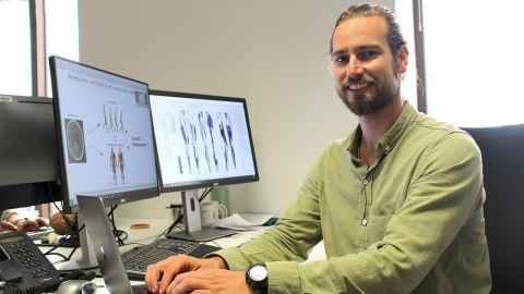
“We have some ideas of how growth is impaired by cerebral palsy, but we've never actually looked at it directly,” says Dr Geoffrey Handsfield of the Musculoskeletal Group at the ABI, who is leading the team from the ABI working with Mātai.
CP is a disorder resulting from a non-progressive brain lesion that occurs during, before or soon after childbirth, which impairs movement of the body and limbs. It is the most common form of physical disability in childhood, with more than 10,000 people affected in New Zealand.
CP is not a neurodegenerative disorder but the result of a single-event brain injury that leads to progressive secondary musculoskeletal conditions that affect both the posture and the movement of children with it. Paradoxically, while the neural injury does not change, the musculoskeletal conditions typically get worse over time.
There are interventions that can improve the development of a the musculoskeletal system of a child with CP, if they are assessed and treated early enough, particularly disciplined and ongoing physiotherapy, as well as orthotics, surgeries and other treatments.
However, how and why the musculoskeletal system degenerates for children with CP remains poorly understood. Gaining a more scientific knowledge of this process has the potential to inform existing treatments and lead to the development of new ones.
While advancements in surgery have helped with the clinical management of children with CP, many of these interventions are temporary, with symptoms potentially returning over time.
“While advancements in surgery and pharmacueticals have helped with the clinical management of children with CP, many of these interventions are temporary, with symptoms potentially returning over time,” says Dr Handsfield.
“To provide treatment and intervention we need to better understand the musculoskeletal conditions of CP and the underlying impairments of skeletal muscle architecture.”
The study will involve conducting MRI at three time points over two years, using top-of-the-line hardware and world-class computational modelling approaches developed at the ABI.
“My team hopes to not just collect precise and high-fidelity data, but to build on these data with the most sophisticated and accurate analysis techniques possible,” says Dr Handsfield. “It will allow us to quantify differences and changes in size, shape and growth over time.”
Understanding differences in the progression and growth trajectory of leg muscles between participants with and without CP will add to understanding of how ageing and growth contributes to walking impairment for people with CP. “This may help us design new treatment and interventions that could help people with the condition walk better, and for longer, and avoid dependence on a wheelchair
Dr Handsfield and his team are hoping to involve 50 participants (children and adolescents) both with and without CP, who will undergo MRI scanning of their leg muscles three times over a period of two years. “Volunteers will get to see a high-tech scanning technology, that few in New Zealand would normally see,” says Dr Handsfield.
“Few of us get a chance to see the inside of our own leg, all their bones and muscles, and the connective tissues that join them. We’ll send our volunteers the file, so they can have a keepsake. And, if they want, frame it and hang it on the wall!”
The CP project, funded by the Robertson Foundation Aotearoa Fellowship, is one of two projects in which Dr Handsfield is collaborating with Mātai, a non-profit medical imaging research and innovation centre, based in Tairāwhiti Gisborne, which aims to enhance the capabilities of magnetic resonance imaging (MRI) through research and development into advanced medical imaging software and machine learning.
“This is a great model for research investment, one that helps ensure that areas outside of cities and commercial centres have research resources and can engage in this kind of medical research,” says Dr Handsfield.
“It’s about developing research expertise, building connections and providing opportunities for the local workforce, researchers and recent graduates in the health sector and outside the major centres.”
Media contact
Margo White I Media adviser
DDI 09 923 5504
Mob 021 926 408
Email margo.white@auckland.ac.nz