Raising our sights: advances in eye research
3 November 2025
Cover story: From creating corneas from stem cells to investigating eye drops to replace reading glasses, Owen Poland explores University of Auckland research that's changing how we see the world.

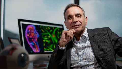
When Professor of Ophthalmology Charles McGhee (ONZM, FRSNZ) arrived at the University of Auckland from Scotland in 1999, things looked very different from how they do today.
“We’ve grown from a subsection of the Department of Surgery, with only one and-a-half senior lecturers, to a standalone Department of Ophthalmology, with 11 professors and associate professors and a total of 89 staff and students,” says Charles.
New Zealand’s first Chair of Ophthalmology, Charles notes that preserving the gift of sight has become a major focus at the University, with researchers in the Aotearoa New Zealand National Eye Centre (ANZ-NEC) trying to address some of the world’s most pervasive problems with potential therapies.
Groundbreaking work includes investigating corneas made with stem cells, and antioxidants that reduce the risk of cataracts.
As a clinician scientist, Charles has participated in more than 550 peer-reviewed publications on research programmes, ranging from corneal transplantation to cataract surgery, the use of stem cells, eye chemical injuries and studies in drug delivery.
Describing cataracts as “a terrible burden” that a developed country like New Zealand shouldn’t have, he initiated the Auckland Cataract Studies and the New Zealand Cataract Risk Stratification project to reduce surgery waiting times. The project has also reduced significant complication rates, which are now less than one percent.
“The risks for a junior trainee surgeon doing elective cataract surgery and a senior surgeon are now the same because we stratify the patient risks,” he says.
Another long-term interest is the diagnosis and treatment of keratoconus (where the cornea thins, bulges and distorts) which is very common among young New Zealanders, particularly Māori and Pacific peoples.
In response, Charles introduced a technique called collagen cross-linking to the New Zealand public health system, which can stop the disease and prevent vision loss by increasing the chemical bonds between the collagen fibres that hold corneal tissue together.
“Probably between 700 and 1,000 cases are now treated each year, and it all largely started with Dr Charlotte Jordan’s PhD project in 2007, which involved the University of Auckland and Auckland District Health Board.”
We’re creating this whole platform of researchers, which includes a growing
number of Māori and Pacific students.
On the clinical side, he’s trained a legion of junior surgeons and performed more than 1,000 corneal transplants and several thousand cataract procedures. He’s also introduced many novel surgical techniques, such as cultured limbal stem cell transplants that regenerate and restore damaged corneas.
And many of those skills are being passed on to a new generation of surgeons at the University’s Calvin Ring microsurgical lab, where hands-on surgery is taught with the aid of two virtual-reality simulators.
“You believe you’re operating inside an eye doing cataract or retinal surgery,” says Charles, “and research confirms this approach consistently reduces complication risk for trainee surgeons early in their clinical career.”
Along the way he’s supervised around 40 PhD students, including Esmeralda Lo Tam – a New Zealand Sāmoan and one of his keratoconus patients – who is now studying the equity of eyecare delivery, including keratoconus treatment, among high school students and in the community (see below).
“We’re also creating this whole platform of researchers, which includes a growing number of Māori and Pacific students,” says Charles.
As a founding director of the ANZ NEC – which includes the departments of Ophthalmology, and Optometry and Vision Science, and the Molecular Vision Laboratory – Charles has brought together more than 200 staff and students under one umbrella.
Peer-reviewed grants, industry collaborations and philanthropy have all played a key role in fostering research. He’s particularly grateful to the Paykel, Hadden and Gray families for their staunch support of major initiatives, such as his Chair of Ophthalmology, and that of Professor Colin Green.
The department, says Charles, is typically seen “as one big family”. “I think that’s probably why we appeal to philanthropy. They see what they’re getting; they see it’s a happy, bustling, fun place to work.”

Unlocking the eye’s inner secrets
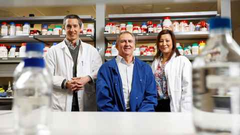
A foundation partner of the ANZ-NEC is the University’s Molecular Vision Research Cluster (MVRC). Founded by the Department of Physiology’s Professor Paul Donaldson, it consists of a multidisciplinary team whose combined expertise provides a rich training environment for the study of eye diseases.
Their groundbreaking cataract research has led to a new understanding of how water and antioxidants flow around the lens within a unique microcirculation system. The work led to a University of Auckland Research Excellence Medal in 2025 for Paul and his team.
“We were one of the first to show how it could potentially be used for new therapies to reduce the incidence of cataract,” he says.
Age-related nuclear cataract is the world’s leading cause of blindness, and diabetes is another factor contributing to rising cataract numbers. Around 30,000 cataract surgeries are done each year in New Zealand, with long wait lists, and the condition is an increasing burden on the health system.
“We’re getting an epidemic because people are living longer and experiencing the effects of cataract,” says Paul. “That’s increased by the fact that we’re also facing a diabetic epidemic with obesity.”
The use of mass spectrometry, to analyse the composition and structure of molecules, has played a key role in helping unlock the inner secrets of the eye. Says Associate Professor Gus Grey: “We’ve been able to show that there are very different spatial regions in the lens and that has a very particular bearing on lens function.”
Supported by a $1.2 million grant from the Health Research Council, Gus will spend the next three years investigating how the uptake and metabolism of glucose in the human lens contributes to diabetic cataract.
“We will be looking at specific proteins and their modifications so that we can better understand how we can develop human-specific treatments to delay the onset of cataract,” says Gus.
We were one of the first to show how it could potentially be used for new
therapies to reduce the incidence of cataract.
Joining Gus and Paul on the project is Associate Professor Julie Lim, another MVRC scientist, whose research into the role of oxidative stress in eye disease has enabled the team to make a crucial advance from animal models to human tissue.
One option being investigated is the potential use of the antioxidant glutathione and its derivatives, although one of the major challenges will be to deliver therapeutic doses of antioxidants to the lens, which doesn’t have a bloodstream.
“A lot of our work has been trying to understand the best ways to deliver an antioxidant to the lens,” says Julie. “It’s not as simple as taking an oral antioxidant supplement and hoping that your cataract will be prevented.”
Julie is also researching why more than 80 percent of patients develop a cataract after having a vitrectomy – a surgical procedure undertaken to treat various conditions affecting the retina. By using state-of-the-art, non-invasive MRI imaging to monitor changes in oxygen levels, she hopes to protect the lens from cataract.
“We’re working closely with our colleagues in the Department of Ophthalmology to design particles that contain antioxidants that lower oxygen levels in the eye and protect the lens from cataract after vitrectomy.”
And while solutions have yet to be devised, says Paul, it could involve controlling water circulation in the eye to deliver a combination therapy of antioxidants to the right targets.
“If we can delay the onset of cataract by five to ten years, we’ll halve the incidence of cataract,” he says, “and that reduction would mean a huge saving in time and resources.”

The gift of sight
Much of the human tissue used for advanced research is provided by the New Zealand National Eye Bank, located within the Department of Ophthalmology.
Established in 1991, the non-profit organisation is dedicated to the prevention of blindness through the provision of donated corneas. These have given more than 8,000 grateful New Zealanders a last chance to save their sight.
“There’s a transplant pretty much every day of the week around the country,” says Nigel Brookes, the eye bank’s technical specialist who has overseen the process since its inception.
In addition to analysing data on what’s been transplanted and why, Nigel also conducts clinical research and has developed a software programme to identify the endothelial cell density of corneas to determine their suitability for transplant.
“We have images of the endothelium before they get transplanted and we’re comparing them to ones that have been transplanted, and we can see the density decreases massively after transplant,” he says.
One of the biggest challenges is a shortage of donors to satisfy a waiting list for around 500 corneal transplants. About 40 percent of the tissue currently being used has come from Australia, and some is also being imported from the US, although “we’re aiming hard to get self-sufficiency again,” says Nigel.
To that end, a new initiative will be starting at Auckland City Hospital to source eye donations from areas outside of ICU and also to educate staff, patients and families about the process. “Everyone knows about heart transplants and kidneys, but eyes are not talked about very much.”
He’s also “deeply confused” about the donor process attached to drivers’ licences, because next of kin can ultimately veto any donations, and he supports having a registry to clarify donor intentions.
Eye bank coordinator Marisa Thi, who spent her early career conducting animal research, says that it’s “incredibly rewarding” being able to help restore people’s sight.
“They’ve been able to drive again, they’ve been able to go back to work, and just enjoy doing all the things that they love doing.”

The promise of stem cells
Just a few steps down the corridor at the department, Professor of Ophthalmology Trevor Sherwin’s team of PhD students and dedicated lab technicians are forever grateful for the donation of tissue to advance their cornea and stem cell research.
“None of this work could happen without those families making the brave decision to donate their loved one’s tissues,” says Trevor.
Using cells from umbilical tissue, his team are looking to regenerate a range of corneal cells to trial in donated cornea. “What we’re trying to do is regenerate the tissues and restore the function again, and that’s the promise of stem cells.”
Funded by the Auckland Medical Research Foundation, the Save Sight Society and the Freemasons Foundation, the research aims to inject stem cells into the cornea, says Trevor, “so that we can recreate the cornea in the patient rather than recreating in a dish and then transplanting it”.
Bioengineering new hybrid material that mimics eye tissue is another area of research; in collaboration with the Faculty of Engineering, PhD candidate Dr Judith Glasson has been extracting crystalline proteins from fish eyes to create corneal implants.
“What we’re trying to do is make implants that are a really good substitute and can be used at much earlier stages, and much more widely than the current tissues,” says Trevor.
The ultimate satisfaction would be to see our eye drop go into the clinic and
restore people’s lives.
Cell reprogramming is yet another research strand, and a start-up company called TheiaNova has been formed on the back of this. It’s developing world-first eye drops to ‘turn back the clock’ and regenerate collagen matrix molecules in people suffering from keratoconus.
“We believe that if we put a contact lens on the cornea while that molecule is being made, then we can reshape the cornea back to being functional.”
Trevor has published more than 100 papers and received more than 6,000 citations, however he says that working with the University’s best and brightest young people provides day-to‑day satisfaction. “And the ultimate satisfaction,” he says, “would be to see our eye drop go into the clinic and restore people’s lives.”
Eye drops instead of reading glasses
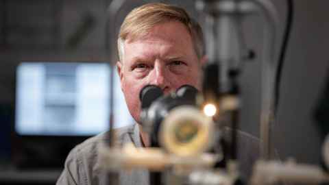
When Dr Alyssa Lie isn’t lecturing at the School of Optometry and Vision Science, or treating glaucoma patients in a private practice, she’s continuing her quest to discover why people develop presbyopia – or farsightedness – and how eye drops could correct it.
“It’s an age-old problem,” she says, “but we’ve not really cracked the code on why it develops or how to reverse it. We just know how to fix the problem and to make the vision clear again.”

Fixing presbyopia has traditionally involved the use of glasses or contact lenses. However, Alyssa says the launch of Vuity (trademarked) eye drops in 2021 provided the “biggest breakthrough in a century” – and an opportunity to advance her research.
Despite the eye drops being FDA approved for prescription-only use to treat presbyopia, there’s “limited evidence”, she says, about how they work – other than making the pupil shrink to increase the depth of focus. “It is an optical trick to make you feel like the book you were reading isn’t very blurry anymore.”
Using MRI scans to observe what effect the eyedrops had on the front part of the eye, Alyssa’s initial clinical trial revealed that the eye drops changed the way water is being distributed inside the lens. “That’s very exciting, because how water is distributed in the lens tissue actually determines the optical power of the lens.”
She is now recruiting volunteers experiencing presbyopia for another clinical trial to test the effect of the eye drops on light-sensitive tissues at back of the eye, like the retina, where any changes could have a profound effect on vision.
The total global costs associated with uncorrected presbyopia have been estimated at US$30.8 billion. Alyssa says the aim would be to create a new drug that could manipulate water distribution in the eye without current drawbacks, such as reduced night vision.
While her previous research was funded by donors including the University of Auckland Research Development Fund, the Maurice and Phyllis Paykel Trust and the US National Institutes of Health, her latest trial is supported by the Freemasons Foundation and the highly influential US Association for Research in Vision and Ophthalmology.
“That is a really big recognition that this is very much a question that needs to be answered,” says Alyssa.

From patient to PhD
Esmeralda Lo Tam was living in Sāmoa when one day she got a sore eye. She rubbed it, rinsed it with water but it only felt worse.
The next day she woke up only able to distinguish light and dark. She arrived at the hospital to be told its sole ophthalmologist was away and would be back in a week.
“At this stage,” recalls the University of Auckland ophthalmology PhD candidate, “the pain was excruciating.”
With all her family back in New Zealand, where she was born and raised, she made the call to catch a midnight flight out and was seen at the Eye Institute in Manukau first thing the next morning.
Staff diagnosed severe keratoconus – a condition where the cornea thins, bulges and distorts – requiring immediate hospitalisation. “They said if I’d waited one day longer in Sāmoa, I would have lost my eye completely.”
A long period of treatment – ultimately a corneal transplant followed by two rejection scares – and recovery followed. There were long hospital stays, and persistent double vision meant she was reliant on others. She tears up recalling the moment her vision was restored: “I remember it vividly. The two pictures I’d been seeing were suddenly one.”
Despite having a masters in public health when diagnosed, Esmeralda had never heard of keratoconus. One day during her treatment, she asked ophthalmologist Professor Charles McGhee why she’d developed the condition, which affects one in 2,000 New Zealanders, but appears to be more common and severe in Māori and among Pacific peoples.
“He said, ‘Actually, a lot of Māori and Pacific people have this condition, and we don’t have any Pacific researchers in this area to be able to understand it on a deeper cultural level’.
“So I said, ‘Can I do it?’”
Charles accepted her offer, and she’s now undertaking her PhD looking at the prevalence of eye diseases in Pacific communities. She’s part of a team screening the vision and eye health of 1,000 young people across 16 Auckland high schools with high levels of Pacific students, with screening of 2,000 Pacific people in the community to follow.
Raised embedded in her Sāmoan language and culture, Esmeralda hopes insights from the research will help catch eye health and vision conditions earlier among Pacific people, avoiding the complex and expensive treatment she underwent.
“I couldn’t imagine I would be studying in public health, but also able to do a little clinical work,” she says of her PhD. “That’s all thanks to Charles and his team, supporting me on that journey, out in our Pacific communities.”
The full Spring 2025 issue of Ingenio includes feature stories on how science is explaining the power of the arts; smart ideas for a more secure energy future; and whether we need a 'right to disconnect'.
Plus, read alumni profiles on gin master Marcel Thompson, DNA detective Bethany Forsythe, and food-personality-turned-novelist Julie Biuso, as well as the latest University news, research, arts coverage and more.