Medical devices and technologies
Mechatronics research focused on providing real-world solutions to surgical, assistive and rehabilitation challenges.

Current projects
Prosthetics
This research focuses on the development of prosthetics using novel material actuators and sensors, to enable more lightweight and dexterous prosthetic devices in the future.
Paediatric gait trainer
In this project, a novel robotic gait trainer (RGT) will be developed to directly improve the walking effectiveness of children with cerebral palsy through administering a range of therapy regimes. The RGT will also act as a tool to help uncover new fundamental knowledge of the factors relating to the development of gait patterns, including individual trends and habits when learning to walk.
Upper limb exoskeletons for children
The robots are developed to manipulate the muscles of a child just as a therapist would, enabling stretching, strengthening and range-of-motion therapy.
Gait rehabilitation for stroke
This research focuses on rehabilitation for people who have experienced a stroke and are now faced with the challenge of re-learning to walk. We are developing robots that can guide the legs of a patient to help them get up and walk again.
Ankle assessment and rehabilitation robot
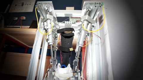
This research focuses on developing an intelligent ankle assessment and rehabilitation robot for both passive and active training. Current work involves the development of a wearable, parallel robot that can be used to assist therapists in the treatment of a variety of ankle injuries and motor rehabilitation. The desired outcome is to have a design that is adaptable to various patients and to identify, develop and implement an intelligent controller for adaptive rehabilitation exercises based on real-time robot-assisted ankle assessment.
Tool-tip tactile and nerve sensing for minimally invasive spinal surgery (MISS)
A MISS surgeon interacts with tissue and spine via long, slim surgical tools through small and deep incisions under image guidance but has no actual sense of direct touch that helps discriminate types of tissues and bones for safe operation. This project proposes to embed a single optic fibre into the surgical tool and use Fabry-Perot interference to measure force and Raman spectroscopy to detect the types of tissues at the tool tip. The project involves laser optic circuit design, signal process, tool redesign and fabrication, laboratory calibration and clinical trials.
Smart medical needle for percutaneous surgeries
A flexible bevel-tip needle has the ability to navigate around obstructions like tumours, organs and nerves and perform surgeries at a targeted location. It can be used for minimally invasive surgeries, including brain, liver and prostate surgeries, blood/fluid sampling, regional anaesthesia and catheter insertion. This project proposes a fibre optic system for measurement of the spatial deflection of the needle and the tip force of the needle interacting with tissue, investigating a sensor-based algorithm to plan optimal motions of the needle and the development of a robotic device to steer the needle to navigate around obstructions by means of percutaneous insertion.
Swallowing robotics for dysphagia management
The aim of this research is to evaluate this novel rheological investigation tool as a model of both healthy and diseased swallowing states and to investigate the interaction between boluses and the oesophagal peristaltic transport process. Videofluoroscopic contrast studies and manometry will be used to measure the swallowing behaviour of the robotic oesophagus. These tools are the gold standard in human dysphagia assessment with widely published normative data. Achieving these specifications will demonstrate the transparency of using the device for medically significant prediction of swallowing. The device exhibits superior control of variables known to have sensitive effects on swallow function throughout experimentation. The quantitative nature of the investigation will reduce dependence on subjective measures and will allow for improved inter-study comparison and definition of rheological terms.
Large deformation peristaltic actuator
Recent research involved the development of an actuator as a robotic instrument that reproduces the movement of internal organs such as the oesophagus, stomach, intestines and heart and can be used to validate clinically significant hypotheses, test the performance of different catheters (sizes, shape, flexibility etc.), and examine the flow of the contents in those organs. The principle of muscular hydrostat and other biological and physiological principles will be followed to specify the design requirements of the actuator. The mechatronic design principles will be applied in developing the bio-robotic actuation system. Novel drives (such as the PneuNet technique) and control methods (such as data-driven learning control) will be developed to make the actuator useful for various medical purposes.
Using bio-signals to control robots
Our motivation is to develop a 'human-centred' physiological controller to incorporate human intention for controlling the human-inspired rehabilitation robots. The biomechanics techniques such as 3D musculoskeletal model, motion analysis technique and muscle force estimation methods are implemented in the physiological controller design. Two models are developed based on the patient-specific musculoskeletal model: the inverse dynamics-static optimisation-based muscle force estimation and the EMG-driven musculoskeletal model. The human-inspired gait rehabilitation robot is the case study that shows the effectiveness of the proposed controller.
Image-guided surgery
Image-guided surgery (IGS) is a real-time link of the operative field to a pre-operative imaging data set (either CT or MRI images). The aim is to create a generic IGS system that can be applied to many surgical procedures. Surgeons can utilise the system to visualise a 3D model of an anatomical structure and carry out any required exploration prior to operation. Later, the system will be able to provide real-time practical aid to surgeons during surgical procedures by shortening operating times and reducing a procedure’s invasiveness, all of which can lead to better patient outcomes and faster recovery.
Brain-computer interface based on steady-state visual evoked potential
This research focuses on the electroencephalograph (EEG) based BCI, and in particular, it uses a visual evoked potential called the steady-state visual evoked potential (SSVEP). In this paradigm, visual stimuli modulated at different frequencies are presented to the user. Each frequency is associated with an action in an output device (e.g., turn left/right in a mobile robot). When the user focuses attention on a certain frequency, the corresponding stimulating frequency appears in the spectral representation of the EEG signals recorded at the occipital sites. These signals are then subject to signal processing, where the relevant signal features are translated into device commands.
Courses
- MECHENG 736: Biomechatronics
- ENGGEN 770: Medical Devices Technology
- ENGGEN 771: Medical Devices Practice
- ENGGEN 791A/B: Medical Devices Research Project
- ENGGEN 793A/B: Medical Devices Research Portfolio