Cardiomyometer
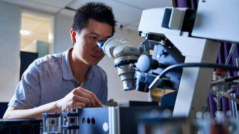
Our cardiomyometer simultaneously measures the geometric, mechanical, energetic, and ionic properties of individual realistically-contracting heart muscles.

With each beat, the heart’s cells release a brief pulse of calcium, which triggers force development and cell shortening. This process consumes energy and oxygen and produces heat. Researchers need to be able to measure all these processes simultaneously, while subjecting heart muscle to realistic contraction patterns that mimic the pressure-volume loops experienced by the heart with each beat.
We have completed the construction of an innovative testing device that achieves this goal: The Cardiomyometer. With this device, we can, for the first time, follow all of these subcellular events, beat by beat, in living heart tissue under either normal or diseased conditions.
How it works

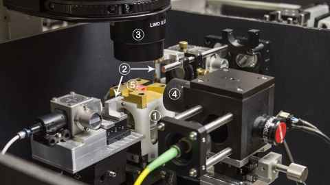
Heart muscle samples, about the diameter of a human hair and about 2 mm in length, are loaded into the instrument sensor (labelled ‘5’) and kept alive by flowing solution. We have developed and integrated into this device several innovative measurement systems:
- Fluorescence imaging system for recording calcium release
- Muscle force and length control apparatus
- Microscope system and algorithms that determines internal tissue movement, strain and sarcomere length
- Optical Coherence Tomography system for measuring 3D shape change, during contraction
- Temperature sensors for measuring muscle heat production.
This system is now being used to study
- The complex relationships between muscle mechanical parameters (force and length), calcium transient dynamics, calcium sensitivity of the myofilaments, and the resulting length distribution and muscle displacement/strain.
- The shortening that occurs inside a muscle sample as it performs work against a “realistic” time-changing load.
Publications
- Dowrick, J. M., Anderson, A. J., Cheuk, M. L., Tran, K., Nielsen, P. M. F., Han, J.-C., & Taberner, A. J. (2021). Simultaneous Brightfield, Fluorescence, and Optical Coherence Tomographic Imaging of Contracting Cardiac Trabeculae Ex Vivo. JoVE(176), e62799.
- Cheuk, M. L., Lam Po Tang, E. J., HajiRassouliha, A., Han, J. C., Nielsen, P. M. F., & Taberner, A. J. (2021). A Method for Markerless Tracking of the Strain Distribution of Actively Contracting Cardiac Muscle Preparations. Experimental Mechanics, 61(1), 95-106.
- Cheuk, M. L., Anderson, A. J., Han, J. C., Lippok, N., Vanholsbeeck, F., Ruddy, B. P., Loiselle, D. S., Nielsen, P. M., & Taberner, A. J. (2017). Four-Dimensional Imaging of Cardiac Trabeculae Contracting In Vitro Using Gated OCT. IEEE Transactions on Biomedical Engineering, 64(1), 218-224.