Changes in lumbar curvature and lumbopelvic pain during pregnancy
A pregnancy-specific model will be developed to estimate lumbopelvic loads during activities, improving understanding of lumbopelvic pain mechanisms in pregnancy.

About this research study
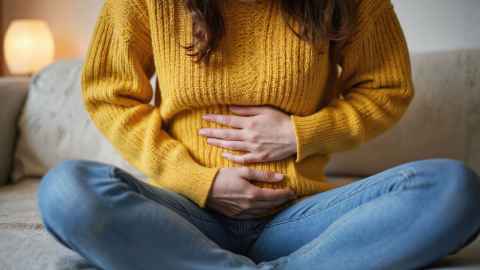
Pregnancy is one of the most common and important events in the life of women. It is usually divided into three stages: the first trimester (weeks 1–13), second trimester (weeks 14–26), and third trimester (week 27 until birth). During pregnancy, the body goes through huge changes in shape, hormones, and physical function. These changes can affect how the body moves and feels, often leading to discomfort, reduced stability, and pain. Pain in the lower back and pelvis region—together called lumbopelvic pain—is the most common physical complaint during pregnancy. This pain usually gets worse as pregnancy progresses, and can affect daily life, work, and sleep. Unfortunately, these issues are frequently underestimated, with many viewing them as a temporary and normal aspect of pregnancy. However, research shows that for some women, the pain can continue after birth or even become long-term.
Many factors may contribute to this pain, such as the growing weight of the womb, increased curve in the lower back (to balance the extra weight), looser joints due to hormones, separation of the pubic bone, weaker muscle control, and changes in walking patterns. Using computer simulations (based on body shape and movement data collected during pregnancy), we can estimate the loads on joints and muscles in the lower back and pelvis during everyday movements. This helps us understand why pain happens and may even help predict how bad the pain will be. Therefore, we need a detailed, pregnancy-specific model to better understand what causes this pain—and to help develop better ways to prevent or treat it.
What's involved?
Participants will attend one session in each trimester (first, second, and third) and one postpartum session. Data collection will include:
- 1x Health checks (i.e. heart rate, temperature and blood pressure)
- 1x 3-dimensional whole-body imaging (non-hazardous)
- 1x Motion capture of body movements during walking and some common daily activities (non-hazardous)
Each session will last approximately 60-90 minutes.
Eligibility criteria
Inclusion criteria:
- Women preparing for pregnancy or pregnant women in the 1st trimester (weeks 1–12)
- Maternal age 20–40 years
- Parity ≤3
- The absence of pre-pregnancy lumbopelvic pain
- The absence of previous lower back or pelvic surgery
- In a normal mental state, has signed an informed consent form and can cooperate with the researcher
Exclusion criteria:
- Those with unstable conditions or other serious systemic diseases. (American college of obstetricians and gynecologists in the guide pointed out that have the following need to rule out: patients with severe lung disease, cervical dysfunction or cerclage, have the risk of a pre-term birth twins or multiples, 26 weeks pregnancy diagnosis of placenta previa, premature delivery or premature rupture of membranes, preeclampsia, gestational hypertension or severe anemia, cardiovascular disease.)
- People with mental illness can't cooperate with those who complete the questionnaire.
- Women with a BMI>32 in their first trimester or before pregnancy are more at risk of developing complications during pregnancy.
What you will receive
$20 voucher for each visit
Participant information
Get involved
If you would like to know more about this study or to apply, we would love to hear from you:
Jie Chen
Research student
Email: jche973@aucklanduni.ac.nz
Dr Julie Choisne
Principal Investigator
Email: j.choisne@auckland.ac.nz
Approved By The Auckland Health Research Ethics Committee On 27/11/2024 For A Period Of 3 Years. Reference Number 27603.
Recruitment ends: 1st June 2026