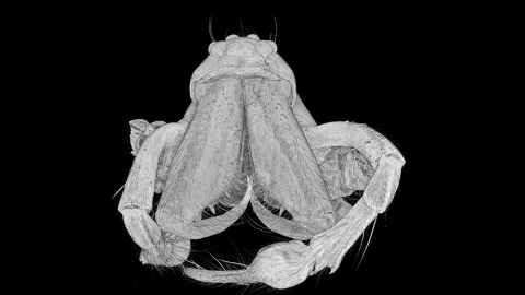
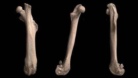
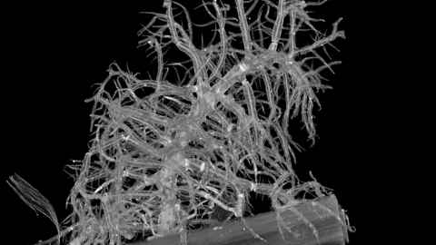
Images and sample requirements


MicroCT is not a quick technique as the acquisition of thousands of individual x-ray projections can take hours per sample.
The time required is highly dependent on sample size, density and spatial resolution requirements.
It is both data-acquisition and computationally intensive so it requires many Tb of storage and high-end computing hardware.
MicroCT facility staff can image your samples for you, or train you to image your own.
Sample requirements
- Able to fit in the MicroCT chamber
- Allows 100 kV X-rays to pass through
- Differing X-ray absorption to provide contrast
- Soft biological tissue requires staining
- Dry or wet samples.
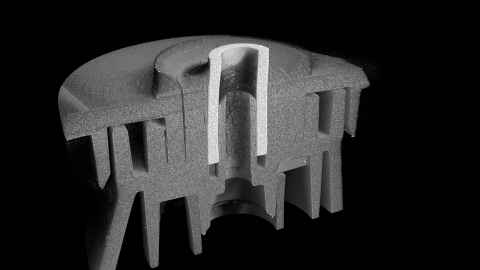
Magnification
Magnification in CT is achieved by the geometrical relationship of the source, sample and camera positions. The instrument magnification range is 1.4x to 14.9x.
Pixel resolution, field of view and sample size are interdependent.
Large sample = low resolution and small sample = high resolution.
Low magnification
27.0 µm pixel resolution. Field of view 27 x 17 mm. Sample diameter 25 mm.
High magnification
0.35 µm pixel resolution. Field of view 1.7 x 1.1 mm. Sample diameter 2 mm.
Capabilities
- MicroCT instruments are able to record multiple image fields – up to three fields wide and five fields high and stitch them together.
- The MicroCT can image samples that are 75 mm wide and 80 mm high as long as x-rays can pass through the sample and give sufficient signal.